
The human heart is a muscular organ—about the size of an adult fist—located slightly left, anterior of the vertebral column, and posterior of the sternum. It is responsible for pumping oxygenated blood to the body and deoxygenated blood to the lungs through continuous rhythmic contractions.
How the heart beats
The myogenic heartbeat originates from the sinoatrial (SA) node—a natural, intrinsic pacemaker—within the heart itself rather than from specific nerve impulses.
Without extrinsic neural (autonomic nervous system) or hormonal influences (endocrine system) the SA node pacing rate would be around 100 beats per minute.
The heart rate and cardiac output, however, must alter under varying circumstances in response to the demands of the body for oxygen and nutrients. The nervous system, hormones, and other factors influence this action.
The average heart rate changes through the lifespan:
- Infant: 120 beats per minute.
- Child: 90 beats per minute.
- Adolescent and adults: 60–100 beats per minute.
Your heart’s electrical system
Autonomic and endocrine control of cardiovascular function
Heart contraction: Diastole and systole
Kids health: Your heart and circulatory system
The science behind compressions
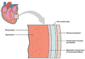
Layers of the heart wall
Heart Valves. (© OpenStax College/Wikimedia Commons/CC-BY-SA-3.0)
A double-membrane sac, the pericardium, encloses three layers of the heart.
- Epicardium: The epicardium is the outer protective layer of the heart that assists in the production of pericardial fluid.
- Myocardium: The myocardium is a muscular middle layer of the heart, which enables the heart to contract.
- Endocardium: The endocardium is the inner layer that lines the heart chambers and covers the heart valves.
Diagnosis and management of pericardial effusion
Exterior of the heart
Diagram of the heart. (© Wapcaplet/Wikimedia Commons/CC-BY-SA-3.0/GFDL)
Several main blood vessels of the circulatory system connect to the heart by its exterior to supply blood to the heart itself (coronary circulation) and to the four chambers inside the heart (left atria, right atria, left ventricle, and right ventricle).
The right atrium receives deoxygenated blood from the inferior vena cava and superior vena cava and then sends it to the right ventricle.
The right ventricle contracts to pump deoxygenated blood from the heart to the lungs, via the pulmonary arteries, to expel carbon dioxide during exhalation and pick up oxygen during inhalation.
From the lungs, through the pulmonary veins, the left atrium receives oxygen-rich blood and pumps it into the left ventricle.
The left ventricle contracts to deliver the oxygenated blood out the aorta to the cells within the body.
- Left anterior descending artery: The left anterior descending artery is the largest coronary artery that supplies blood to the heart. It is located behind the pulmonary artery.
- Right coronary artery: The right coronary artery is an artery that runs toward the heart’s crux (the area on the lower backside of the heart) and primarily supplies blood to the right side of the heart.
- Left coronary artery: It rises out of the aorta above the aortic valve’s left cusp to feed blood to the left side of the heart.
- Superior vena cava: The superior vena cava is a vein that carries deoxygenated blood from the upper body into the right atrium.
- Inferior vena cava: The inferior vena cava is a large vein carrying deoxygenated blood from the lower body into the right atrium.
- Pulmonary arteries: The main pulmonary artery splits into the right and the left main pulmonary artery. The pulmonary arteries carry oxygen-depleted blood into each corresponding lung. All arteries, except for pulmonary arteries, transport oxygen-rich blood.
- Pulmonary veins: The left and right pulmonary veins—two from each lung—carry oxygen-rich blood from the lungs into the left atrium. The pulmonary veins are the only major veins in the human body that carry oxygen-rich blood.
- Aorta: The aorta is an artery that sends oxygen-rich blood out to the cells of the body.
Classification and structure of blood vessels
Accurate definition of the widowmaker heart attack
Fetal Circulation – The umbilical vein supplies the fetus with oxygen-rich blood from the placenta and the fetal heart pumps oxygen-depleted blood via the umbilical arteries back to the placenta.
Right side of the heart
The right side of the heart—the right atrium—is where the largest veins, the inferior vena cava, and superior vena cava, return deoxygenated blood from the lower and upper parts of the body.
From these veins, the blood travels into the right atrium and is pumped into the right ventricle.
From the right ventricle, the blood is then pumped through the pulmonary arteries into the lungs.
- Right atrium: The right atrium is a chamber of the heart that receives deoxygenated blood from both the inferior and the superior vena cava.
- Right ventricle: The right ventricle is a chamber of the heart that receives deoxygenated blood from the right atrium.
Reference ranges for normal cardiac chamber size
Double outlet right ventricle (DORV)
Left side of the heart
The left side of the heart—the left atrium—provides a space to receive oxygen-rich blood from the lungs. The blood travels from the lungs, through the pulmonary veins, and then into the left ventricle.
From the left ventricle, the oxygenated blood is ejected out the aorta to circulate within the rest of the body.
- Left atrium: The left atrium receives oxygen-rich blood from the lungs via the pulmonary veins.
- Left ventricle: The left ventricle is a chamber of the heart that receives oxygenated blood from the left atrium.
Left atrium and pulmonary veins
Understanding heart function: What is the ejection fraction?
Interior of the heart
The interior of the heart helps maintain the life-sustaining flow of blood. A wall of tissue, known as the septum divides the heart into two halves—this prevents oxygen-rich blood and oxygen-depleted blood from mixing.
- Interatrial septum: The interatrial septum separates the left atria and the right atria.
- Interventricular septum (ventricular septum): The interventricular septum divides the right ventricle and left ventricle.
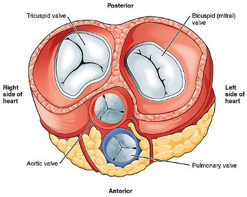
During the cardiac cycle, blood passes through valves that act as one-way inlets for blood flowing into a ventricle and one-way outlets for the blood leaving a ventricle. The Four valves of the heart open and close at precisely synchronized times during one heartbeat.
Heart valves. (© OpenStax College/Wikimedia Commons/CC-BY-SA-3.0)
- Tricuspid valve (atrioventricular valve): The tricuspid valve contains three leaflets and is in between the right atrium and right ventricle.
- Pulmonic valve (semilunar valve): The pulmonic valve is in between the right ventricle and the pulmonary artery.
- Mitral valve (atrioventricular valve): The mitral valve is a bicuspid valve (two leaflets) in between the left atrium and left ventricle.
- Aortic valve (semilunar valve): The aortic valve is between the left ventricle and the aorta.


.svg)